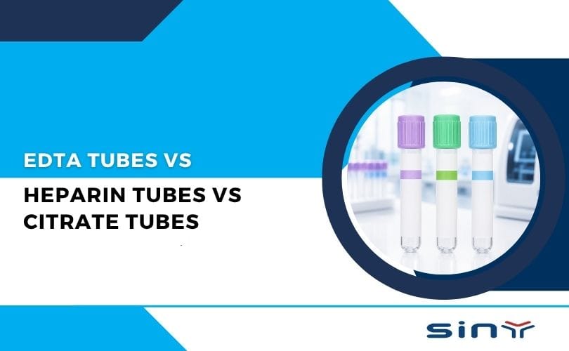
EDTA (ethylenediaminetetraacetic acid) blood collection tubes—often called purple-top tubes—contain EDTA anticoagulant coated on their inner walls. When blood enters the tube, EDTA immediately binds calcium ions (Ca²⁺), halting the clotting cascade and preserving the sample in a true whole-blood state. Clinical laboratories rely on EDTA tubes for a range of hematology tests, including complete blood counts (CBC), manual blood smears, differential white blood cell counts, reticulocyte counts, and glycated hemoglobin (HbA1c) measurements. EDTA preserves red blood cell (RBC) and platelet morphology better than most other anticoagulants, making it the gold standard for accurate cell counts and morphological analysis.
Manufacturers supply EDTA Tubes in two primary salt forms: EDTA-K₂ and EDTA-K₃. EDTA-K₂ dissolves readily in blood and maintains a pH closer to physiological levels, which minimizes RBC shrinkage. Laboratories generally prefer EDTA-K₂ for routine hematology because it yields more stable cell volumes. In contrast, excessive EDTA-K₃—or EDTA-K₃ stored improperly—can cause mild RBC crenation and subtle shifts in mean cell volume (MCV), potentially skewing results.
Anticoagulant Mechanism and Optimal Ratio
How EDTA Prevents Clot Formation
When EDTA Tubes enters blood, it rapidly chelates free Ca²⁺ ions, a vital cofactor in the coagulation cascade. By binding calcium, EDTA interrupts the conversion of prothrombin to thrombin and prevents fibrinogen from polymerizing into fibrin. This immediate chelation keeps the sample in a liquid state, ensuring accurate cell counts and morphology assessments.

EDTA Concentration vs. Blood Volume
Standard Ratio: Industry guidelines specify roughly 1.5–2.2 mg of EDTA per milliliter of blood. In practice, a 2 mL tube contains about 20 µL of EDTA-K₂ solution at approximately 180 g/L concentration. This ratio ensures effective anticoagulation without over-dilution.
Underfilling Risks: If you underfill the tube, too much EDTA relative to blood volume dilutes cellular elements and lowers observed counts. Underfilling can also alter measured hematocrit and hemoglobin values.
Overfilling Risks: Overfilling depletes EDTA too quickly, allowing microclots or cell aggregation. Even small clots can trap cells, leading to falsely low counts and abnormal cell morphologies.
Filling Target: Aim for at least 50% of the tube’s nominal volume, ideally matching the marked fill line (2 mL or 3 mL). That balance maintains anticoagulant efficacy and preserves cell integrity.
Storage Conditions and Shelf Life
Prepared EDTA Solution: Once you make the EDTA solution, store it at 0–4 °C and use it within seven days. Over time, EDTA degrades and loses chelating power.
Sealed EDTA Tubes: Keep unopened EDTA tubes in a cool, dark place. Avoid exposure to temperatures above 25 °C or direct sunlight, which can compromise vacuum pressure and degrade EDTA.
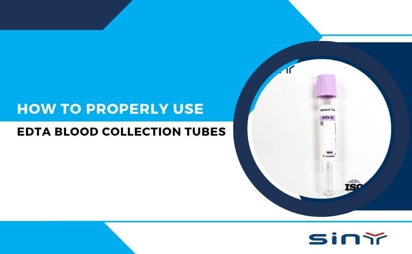
What Are EDTA Blood Collection Tubes and Why Are They Purple?
EDTA stands for Ethylenediaminetetraacetic acid—yeah, it’s a mouthful. But all you really need to know is that EDTA is an anticoagulant, which means it stops blood from clotting. This makes it super useful for tests where the shape and count of blood cells matter—like CBC (Complete Blood Count), blood smears, and even certain types of DNA or molecular testing.
The reason they come with a purple cap? That’s just a universal color code to show that EDTA is the additive inside the tube. According to this detailed breakdown, the purple-top tubes are easily recognizable for fast identification and minimal sample confusion.
Want a closer look? Check out EDTA Tubes for Blood Collection here on EDTA Tube’s official store.
Pre-Collection Preparation
Verify Patient Information and Testing Orders
- Confirm the patient’s identity by comparing their wristband or ID with the lab requisition.
- Ensure you understand which assay labs will perform—CBC, HbA1c, blood typing, or other hematology panels—and that EDTA tubes suit all ordered tests.
Gather and Inspect Supplies
- Use a sterile, single-use venipuncture needle (21–23 G) attached to a vacuum holder compatible with EDTA tubes.
- Prepare 75% alcohol swabs, sterile gauze, a tourniquet, EDTA tubes with intact vacuum seals, adhesive labels, and a rack for upright storage.
- Check each tube’s expiration date. An expired tube may have insufficient vacuum or degraded EDTA, risking clotting.
Skin Antisepsis and Patience
- Scrub the venipuncture site by moving the alcohol swab in concentric circles from the center outward, covering a ~2 cm radius.
- Allow skin to air‐dry completely before needle insertion. Alcohol residue mixing with blood can cause hemolysis or chemical alterations in analytes.
- Label each EDTA tube before drawing blood, writing the patient name, date, time, and “EDTA Anticoagulant” on the side to avoid post-collection mix-ups.
Hand-Held Collection Technique
Apply Tourniquet and Find a Vein
Place the tourniquet 7–10 cm above the antecubital crease. Ask the patient to make a fist, then palpate for a prominent vein.
Reassure the patient and ask them to relax their arm once they see a suitable vein.
Venipuncture and Tube Attachment
Insert the needle bevel-up at a 15°–30° angle. Advance gently until you observe a “flashback” of blood in the hub.
Immediately attach the EDTA tube to the back end of the needle holder. The tube’s vacuum will draw blood automatically. Do not jiggle or force the tube.
Monitor Fill Volume
As soon as blood volume reaches the tube’s marked fill line (2 mL or 3 mL), remove the tube by pushing the sleeve back on the needle holder. Do not pull the tube forcefully; that can break the vacuum seal, leading to underfilling.
Release the tourniquet once blood flows freely. Prolonged tourniquet time can cause hemoconcentration, artificially elevating hematocrit and some analytes.
Immediate Tube Inversion
Within 30 seconds of collection, invert the tube 8–10 times. Hold the tube by the cap and invert it gently in a smooth, back-and-forth motion. This ensures EDTA disperses evenly and prevents clot formation.
Avoid vigorous shaking—the mechanical stress can rupture RBC membranes (hemolysis) or fragment platelets, leading to artificially low platelet counts.
Needle Removal and Hemostasis
Withdraw the needle smoothly, apply sterile gauze pressure to the site for 2–3 minutes, and secure with tape or a bandage once the bleeding stops. Prolonged pressure prevents hematoma formation.
Proper Steps for Using EDTA Blood Collection Tubes
Selecting the Right Tube Size
- Smaller tubes (2-3 mL) are ideal for pediatric patients.
- Standard tubes (4-5 mL) are used for most adult blood draws.
- Larger tubes (10 mL) may be needed for specialized tests.
Correct Order of Draw
EDTA tubes should be drawn after serum tubes (e.g., red-top tubes) but before coagulation tubes (e.g., blue-top citrate tubes) to avoid cross-contamination.
Filling the Tube to the Mark
- Underfilling can lead to excess EDTA, causing cell shrinkage.
- Overfilling may result in insufficient anticoagulant, leading to clotting.
Proper Mixing Technique
- Gently invert the tube 8-10 times immediately after collection to ensure proper mixing.
- Avoid vigorous shaking, as it can damage blood cells.
Storage and Transport
- Store at room temperature (20-25°C) if testing within 24 hours.
- For delayed processing, refrigerate at 2-8°C (but avoid freezing).
For a visual guide on proper blood collection techniques, visit SinyMedical’s YouTube Channel.
EDTA: Advantages and Limitations
Advantages
Superior Cell Preservation: EDTA minimizes RBC crenation and maintains white cell and platelet morphology. That accuracy proves vital when technicians perform manual differentials or when automated analyzers calculate counts.
Rapid, Durable Anticoagulation: A single inversion mix halts clotting immediately, reducing the risk of microclots or platelet clumping. Labs achieve consistent CBC results and accurate platelet enumeration.
Broad Applicability: Besides CBC and morphology, EDTA tubes suit many immunohematology assays, blood typing, and certain molecular tests (e.g., PCR from whole blood).
Limitations
Electrolyte Interference: EDTA chelation lowers measurable Ca²⁺ and Mg²⁺ and can raise K⁺ artificially. For accurate electrolyte panels, use separate tubes (e.g., heparin tubes).
Not Suitable for All Biochemical Tests: Certain assays—like enzyme activities (AST, ALT) or coagulation studies—need native calcium or magnesium. EDTA undermines those tests, requiring other anticoagulants (e.g., sodium citrate, heparin).
Shelf-Life Constraints: Once opened, EDTA tubes lose vacuum and anticoagulant potency over time. Using expired or improperly stored tubes leads to clot formation and test failure.
Summary
Using EDTA blood collection tubes correctly guarantees precise, reliable hematology results. From patient identification and skin antisepsis to proper venipuncture, immediate mixing, correct storage, and timely testing, each step upholds sample integrity. By following these guidelines, clinicians and laboratory staff can minimize pre-analytical variability, avoid sample rejection, and provide physicians with accurate data for patient care decisions. Accurate blood cell counts and morphologies ultimately translate to better diagnostics and improved patient outcomes.
FAQs
Why Choose EDTA Over Other Anticoagulants?
A: EDTA preserves cell morphology best. Heparin can cause platelet clumping; citrate dilutes the sample; oxalate distorts cell shapes. EDTA offers the greatest reliability for CBC, manual smears, and differential counts.
How Soon Must I Run Tests After Drawing?
Room Temperature (18–25 °C): Analyze within 1 hour to prevent cell lysis, platelet clumping, or morphological changes. Refrigerated (2–8 °C): You can extend testing up to 12–24 hours. Beyond that, RBCs begin to leak potassium, platelets lose activity, and white cell morphology degrades, compromising results.
What’s the Best Way to Mix the Tube?
A: Invert the tube 8–10 times within 30 seconds of collection. Perform gentle, smooth inversions—avoid vigorous shaking. Insufficient mixing causes microclots; over-mixing damages cell membranes and generates air bubbles, leading to false counts.
What Happens If I Underfill or Overfill the Tube?
Underfilled: Excess EDTA relative to blood volume dilutes cells, lowering hemoglobin, hematocrit, and cell counts. Overfilled: Insufficient EDTA allows micro clots, causing cell aggregation and falsely low cell counts. Always fill to the tube’s fill line—neither below 50% nor above 110% of nominal volume.
Can I Place the Tube Directly on Ice Packs?
A: Never let a frozen pack touch the tube. Wrap cold packs in cloth or paper and place them beside the tubes in the transport box. Direct contact with ice can freeze cells, causing hemolysis and invalidating results.
Which Form: EDTA-K₂ or EDTA-K₃?
Use EDTA-K₂ whenever possible. Its higher solubility and more physiologic pH minimize RBC crenation and ensure stable MCV readings. Reserve EDTA-K₃ only when EDTA-K₂ isn’t available, but monitor storage conditions closely to prevent concentration changes.