Every blood sample tells a story about a patient’s health. However, that story is only accurate when the sample is collected, mixed, and handled correctly. In modern laboratories, EDTA tubes are among the most commonly used blood collection tubes because they preserve blood cells and prevent clotting. They are essential for Complete Blood Count (CBC) testing and many other hematology procedures.
Despite their widespread use, one critical step is often underestimated: proper mixing immediately after blood collection. Understanding how to mix EDTA tube specimens correctly can make the difference between reliable laboratory results and inaccurate data that may affect patient care.
Many pre-analytical laboratory errors occur before a sample even reaches an analyzer. Improper handling, delayed mixing, and incorrect transport can all compromise specimen quality. While these mistakes may seem minor, they can lead to clot formation, inaccurate cell counts, repeat blood draws, and delayed diagnoses.
This comprehensive guide explains how to mix EDTA tube samples properly after blood draw, why correct mixing matters, common mistakes to avoid, and best practices healthcare professionals should follow to maintain sample integrity.
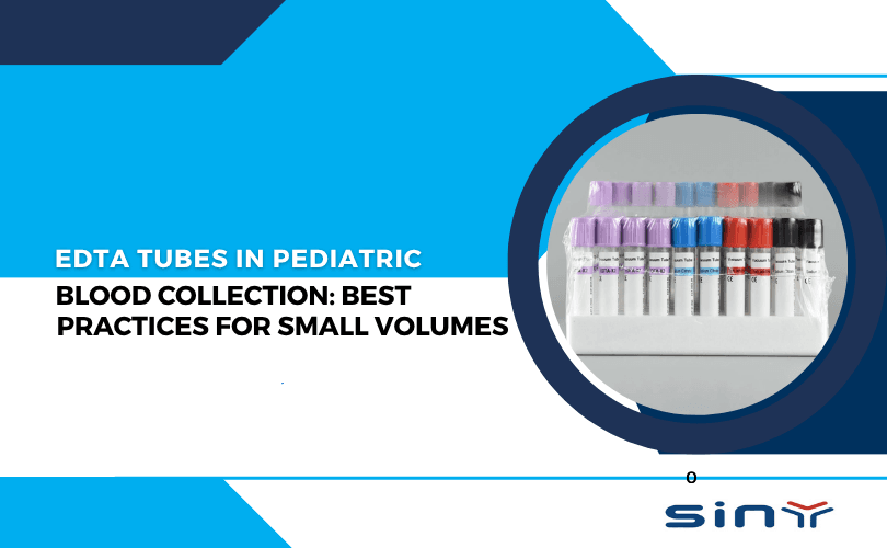
What Is an EDTA Tube and Why Is It Important?
An EDTA tube is a blood collection tube that contains Ethylenediaminetetraacetic Acid (EDTA), a powerful anticoagulant. EDTA works by binding calcium ions in the blood. Since calcium is necessary for the clotting process, removing it prevents blood from clotting after collection.
Because EDTA preserves blood cell morphology exceptionally well, it has become the preferred anticoagulant for hematology testing. Laboratories around the world rely on EDTA tubes for Complete Blood Count analysis, blood smear examinations, hemoglobin testing, hematocrit measurements, flow cytometry, and numerous research applications.
Healthcare facilities looking for high-quality blood collection solutions can explore the main resource center at edta tubeand review available products through products. A detailed range of collection tubes is also available at product category edta tube.
The effectiveness of EDTA, however, depends heavily on proper specimen handling. Even the best tube cannot prevent errors caused by inadequate mixing after collection.

Why Learning How to Mix EDTA Tube Samples Correctly Matters
Many healthcare professionals focus primarily on the blood draw itself while paying less attention to what happens immediately afterward. In reality, the moments following collection are just as important as the venipuncture procedure.
When blood enters an EDTA tube, the anticoagulant coating inside the tube must quickly come into contact with the entire specimen. If the blood remains stationary, portions of the sample may begin to clot before the EDTA has a chance to distribute evenly.
This is why learning how to mix EDTA tube specimens correctly is considered a fundamental part of quality blood collection practice.
Laboratory studies consistently show that pre-analytical errors account for the majority of specimen-related problems. A significant percentage of these issues can be traced back to improper tube handling and inadequate mixing. When mixing is performed correctly, blood cells remain suspended uniformly throughout the sample, helping laboratory instruments generate reliable results.
For patients, this means fewer repeat blood draws and more accurate diagnoses. For laboratories, it means improved efficiency and reduced specimen rejection rates.
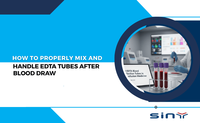
How to Mix EDTA Tube Correctly After Blood Draw
The recommended procedure for how to mix EDTA tube samples is straightforward but must be performed correctly.
Immediately after the tube has been filled, it should be removed from the holder and gently inverted eight to ten times. Each inversion should turn the tube completely upside down before returning it to its original position. The motion should be smooth and controlled rather than fast or forceful.
The goal is to allow the blood to flow from one end of the tube to the other during each inversion. This movement ensures that the EDTA additive distributes evenly throughout the specimen.
One common misconception is that vigorous shaking improves mixing. In reality, shaking can damage blood cells and compromise specimen quality. Gentle inversion remains the universally recommended technique because it achieves thorough anticoagulant distribution without causing cellular injury.
Healthcare professionals who consistently follow this practice significantly reduce the risk of clot formation and specimen rejection.
What Happens Inside the Tube During Mixing?
Understanding the science behind proper mixing helps explain why this simple step is so important.
When blood first enters the tube, the EDTA additive is present on the inner surface of the collection container. Without adequate movement, only a portion of the blood comes into contact with the anticoagulant immediately.
As the tube is inverted, blood repeatedly washes across the tube walls, allowing the EDTA to dissolve and distribute evenly throughout the specimen. This process stabilizes the sample and prevents the clotting cascade from beginning.
Because blood contains millions of cells per microliter, even small inconsistencies in anticoagulant distribution can affect laboratory measurements. Proper inversion ensures that every part of the specimen receives equal exposure to the additive.
The Consequences of Improper Mixing
Improper mixing can create several problems that may not be visible to the naked eye.
One of the most common issues is microclot formation. These tiny clots often develop when blood is not exposed evenly to the anticoagulant. Although they may be difficult to detect during routine inspection, they can interfere with automated analyzers and produce misleading results.
Platelet clumping is another frequent consequence. When platelets aggregate together, laboratory instruments may report falsely low platelet counts. This can potentially lead clinicians to suspect thrombocytopenia when the patient’s platelet count is actually normal.
White blood cell counts may also become unreliable. Since many clinical decisions rely on accurate hematology results, even small inaccuracies can affect patient management.
In severe cases, visible clots form inside the tube, forcing laboratories to reject the specimen entirely. The patient must then undergo another blood draw, increasing inconvenience and delaying diagnosis.
Why Shaking an EDTA Tube Is a Bad Practice
Many healthcare workers mistakenly assume that vigorous shaking improves anticoagulant distribution. Although this belief is understandable, it is not supported by laboratory science.
Blood cells are delicate structures. Excessive mechanical force can damage cell membranes, causing hemolysis. When red blood cells rupture, intracellular contents leak into the plasma, altering test results and potentially making the specimen unsuitable for analysis.
Aggressive shaking can also activate platelets and distort normal cellular morphology. Since hematology testing relies on evaluating intact blood cells, preserving their structure is essential.
Therefore, when discussing how to mix EDTA tube specimens, laboratory guidelines consistently emphasize gentle inversion rather than shaking.
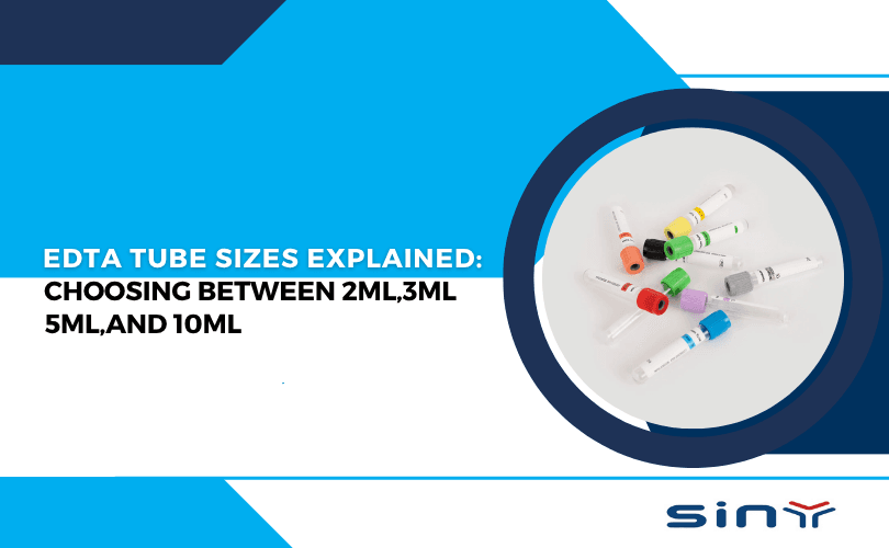
The Relationship Between Tube Fill Volume and Proper Mixing
Another often-overlooked factor is fill volume.
EDTA tubes are manufactured with a carefully calculated amount of anticoagulant designed for a specific blood volume. When tubes are underfilled, the concentration of EDTA becomes disproportionately high relative to the blood sample. This imbalance may cause red blood cells to shrink and can affect certain laboratory measurements.
Conversely, overfilled tubes may contain insufficient anticoagulant to prevent clot formation effectively.
Proper mixing cannot fully compensate for incorrect fill volume. For this reason, healthcare professionals should always follow manufacturer recommendations regarding collection volume.
Those interested in selecting the appropriate tube size can review resources such as how to select the right edta tube size for your
How Proper Mixing Supports Accurate CBC Testing
Complete Blood Count testing remains one of the most frequently ordered laboratory procedures worldwide. According to information available through the Complete Blood Count overview at Complete blood count, CBC testing provides valuable information about red blood cells, white blood cells, hemoglobin, hematocrit, and platelets.
Because CBC analysis depends on accurate cellular measurements, specimen quality directly influences result reliability.
Proper mixing helps maintain uniform cell distribution throughout the sample. This ensures that the portion of blood analyzed by laboratory instruments accurately represents the entire specimen.
Additional information about CBC testing and EDTA tube applications can be found atedta tubes in hematology for cbc tests
Best Practices for Handling EDTA Tubes After Mixing
Proper specimen handling extends beyond the mixing process itself.
Once the sample has been mixed, it should be stored and transported carefully to preserve its quality. Tubes should remain upright whenever possible to minimize unnecessary agitation. Excessive vibration during transport can affect sensitive specimens and should be avoided.
Temperature control is equally important. Exposure to excessive heat can alter blood cell morphology and affect laboratory measurements. Samples should therefore be transported according to established laboratory protocols.
Timely delivery to the laboratory also contributes to specimen integrity. Prolonged delays between collection and analysis may result in cellular degradation, reducing the accuracy of hematology results.
By combining proper mixing with appropriate storage and transport practices, healthcare providers can maximize specimen quality and diagnostic reliability.
Mistakes That Affect EDTA Sample Quality
Even experienced professionals occasionally develop habits that compromise specimen quality.
One common mistake is delaying inversion after collection. Some collectors place the tube on a tray and intend to mix it later. Unfortunately, clotting can begin almost immediately, making prompt mixing essential.
Another frequent error involves incomplete inversions. Tilting the tube slightly does not provide adequate anticoagulant distribution. Each inversion must be complete to ensure effective mixing.
Incorrect tube selection can also create problems. Using a tube size that does not match the required blood volume may affect the anticoagulant-to-blood ratio. Resources such as choosing between 2ml 3ml 4ml and 5ml edta and edta tube sizes explained 2ml 3ml 4ml 5ml 6ml 10 can help healthcare facilities choose appropriate tube sizes.
Finally, rough transportation remains a common source of pre-analytical errors. Even a properly mixed sample can be compromised by poor handling during transit.
The Future of EDTA Blood Collection Technology
Advances in blood collection technology continue to improve specimen quality and laboratory efficiency.
Manufacturers are developing enhanced additive coatings that promote faster anticoagulant distribution. Improved vacuum systems help ensure accurate fill volumes, while new materials provide better specimen stability during storage and transport.
These innovations aim to reduce pre-analytical errors and support increasingly sophisticated diagnostic testing.
Readers interested in emerging developments can explore future of edta blood collection tubes 2026.
Additional industry information is available through edta tube, sinymedical.en.made in china, and educational content published at youtube@sinymedical.
Summary
How to mix EDTA tube specimens correctly is one of the simplest yet most important skills in laboratory medicine. Although the process takes only a few seconds, its impact on diagnostic accuracy is significant. Proper mixing ensures even anticoagulant distribution, prevents clot formation, preserves blood cell morphology, and supports reliable hematology testing.
By immediately performing eight to ten gentle inversions after blood collection, healthcare professionals can reduce specimen rejection rates, improve laboratory efficiency, and help ensure accurate patient diagnoses. When combined with proper tube selection, careful transportation, and timely processing, correct EDTA tube handling becomes a cornerstone of high-quality laboratory practice. Get for more information visit edta tube and contact us.
FAQs
Q: How many inversions are recommended when learning how to mix EDTA tube samples?
A: Most laboratory guidelines recommend eight to ten gentle inversions immediately after collection. This approach ensures proper anticoagulant distribution while minimizing the risk of cellular damage.
Q: Why is gentle inversion preferred when learning how to mix EDTA tube specimens?
A: Gentle inversion distributes the EDTA additive evenly throughout the blood sample without damaging blood cells. Vigorous shaking may cause hemolysis and negatively affect laboratory results.
Q: Can improper handling affect how to mix EDTA tube effectiveness?
A: Yes. Even if the tube is mixed correctly, poor transportation, excessive heat exposure, or delayed processing can compromise specimen quality and influence test accuracy.
Q: Does tube size matter when deciding how to mix EDTA tube samples?
A: The mixing technique remains the same regardless of tube size. However, proper fill volume is essential because EDTA tubes are designed for specific blood-to-additive ratios.
Q: What happens if healthcare workers do not follow proper how to mix EDTA tube procedures?
A: Failure to mix correctly can lead to clot formation, platelet aggregation, inaccurate cell counts, specimen rejection, and repeat blood collection procedures.